Symmastia is caused by over-dissection or oversized implants.
Many people in the breast augmentation field consider a deep, pronounced cleavage as the ideal result that patients seek. Women even bring pictures of models in push-up bras during their first meeting with a plastic surgeon and say that they want their implants to be placed “as close together as possible,” so that they can have that look permanently. Nevertheless, a very subtle and highly perilous borderline exists between tight cleavage and the complication of symmastia that is often overlooked. Symmastia, which is also called “uniboob” or “bread-loafing,” is an extreme disruption of the breast edges. The two separate breasts are no longer separated by a flat sternum but the pockets unite, the skin on the breastbone lifts, and the chest becomes a single continuous mound of tissue.
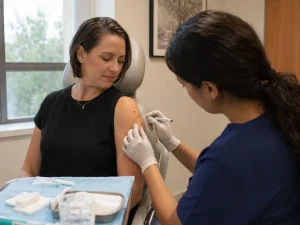
The reason for “What causes symmastia?” is quite straightforward in that it is usually due to surgical mistake involving over-dissection or using an incorrect implant size. It is hardly ever a sudden biological accident but more of a natural failure of the body’s internal midline anchor. The implants will be able to move to the center if the tissues that support the skin to the breastbone are cut. At Lin Europe Clinic, we consider the treatment of symmastia as one of the most difficult plastic surgery complications that have to be fixed, and that is why our main focus is on prevention through accurate measurements and respecting the anatomical “no-fly zone” of the sternum.
The Anatomy of the “Uniboob”
Understanding why symmastia takes place requires some knowledge about the sternum (breastbone). A naturally healthy chest is such that the skin at the center is tightly bonded to the bone beneath it by strong, fibrous connective tissue. Such an attachment acts as a separation, providing the required “valley” or flat area between the breasts. It makes sure that even when you push your breasts together with your hands, they still remain two separate parts that are located in different places.
Symmastia is the result of this important attachment being either completely cut or gradually worn out. If during surgery, a surgeon dissects the internal pocket excessively towards the middle in an attempt to make the implants closer by, the tissue that anchors the skin to the bone will be destroyed. When this skin-to-bone anchor is missing, the skin will tent up as if it is stretched like a drumhead over the chest. The implants, lacking a medial wall, move toward the center and touch one another. The result is a loss of definition; it becomes impossible to floss a bra wire between the breasts as if the skin were floating above the chest wall.
The Culprit: Over-Dissection and Sizing
Implant selection is the other side of the story! Surgical skill, while critical, is just one half of the equation that leads to this problem. Each chest of ours has a certain maximum volume that is primarily determined by its width. Suppose the patient’s chest is narrow with a base diameter of 11 centimeters; however, she demands a 13-centimeter implant to get more volume. This extra width must be accommodated somewhere. The implant physically opens the pockets by pushing them; most of the time, it is the center that gets encroached on.
This is often referred to as the “Cleavage Trap.” Patients who want their implants to touch in the middle intentionally put themselves at the highest risk. Even if the doctor initially leaves a thin strip of tissue, the constant inward pressure from oversized implants can lead to “pressure necrosis.” Over several months or years, this pressure gradually destroys the tissues separating the pockets, causing delayed symmastia that emerges long after the initial recovery period.
Congenital vs. Acquired Symmastia
One of the very few instances where Congenital Symmastia occurs is in women who are naturally predisposed to it. Usually, this means that the breasts are connected by a web of fat and fibrous tissue, which makes the breasts appear webbed. However, the majority of the cases we deal with at our clinic are Iatrogenic, suggesting that the condition was resulted from a previous surgery. Regardless of whether the deformity is congenital or acquired, the basic issue is the same: there is no defined “stop point” for the breast tissue at the middle of the chest.
The Repair Challenge
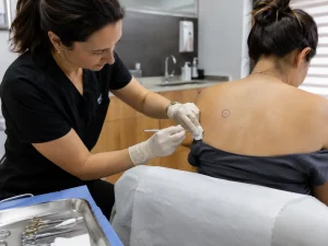
Repairing symmastia is far more complicated than preventing it, and it is regarded as one of the most challenging revision surgeries to be done successfully. It is not possible to “push” the implants apart again. The repair necessitates a detailed reconstruction of the wall that had been destroyed. Internal stitches are generally employed in a procedure called capsulorrhaphy that attaches the scar capsule to the breastbone.
Where there is severe damage, it is a different matter with using threads alone, because the tissue is not sufficiently strong to make them hold. We could have to take a piece of Acellular Dermal Matrix (ADM) or biological mesh to serve as a patch, strengthening the center wall as if it is the internal framework. This patch supports the load and aids in the prevention of the tearing of the stitches. In addition, patients must almost always remodel their implants or downsize to a narrower profile during such a repair to lessen the tension on the new internal wall, hence allowing sufficient time for its healing without being forcibly opened again.
Prevention in Turkey
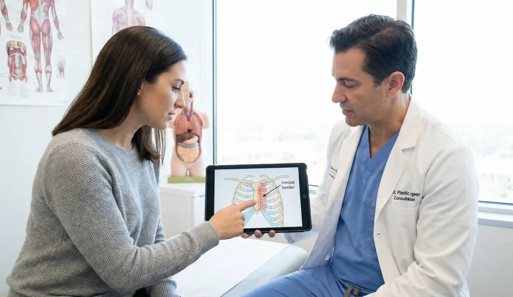
At Lin Europe Clinic in Turkey, a “High Five” Rule (or a comparable geometric principle) is adhered to in the course of every consultation. After measuring your chest width, we deduct the safety margin meant for the sternum in order to arrive at the implant width that can be safely used. We will never place an implant that breaches this safety zone, no matter how much a patient craves “touching cleavage,” because we understand that the long-term consequence is deformity.
We know that a naturally good cleavage is a product of a good bra, not of surgically fused breasts. Our surgeons employ specialized illuminated retractors that let them see exactly where the pocket ends and thus enable them to preserve the vital fibers that make the breasts stay separate and well defined. Preventing symmastia is the only real cure.
Frequently Asked Questions About Symmastia
No, although after repair surgery special compression garments (such as a “thimble”) might be used to support the stitches, no external bra can permanently re-attach the skin to the bone once the internal pocket has merged.
It’s not necessarily the case; although it might be visible instantly if the cutting was very aggressive, it is more common for it to develop gradually over several months as the oversized implants wear away the thin tissue between the pockets.
It is not an emergency health condition and does not have any impact on your heart or lungs, however, it is a serious cosmetic defect that makes it impossible to find properly fitting bras and can cause psychological distress.
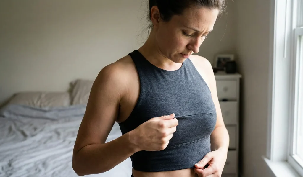
One of the tests you can do at home is the “Pinch Test” where you try to pinch the skin over your breastbone; if the skin is tight to the bone then you don’t have symmastia, but if you are able to lift a “tent” of loose skin away from the bone it means the pockets may have merged.
It is out of the question, since a fractional center fat increase would probably add to the problem, by increasing volume at a location which is supposed to be flat; hence, the issue is a structural loss of attachment rather than a shortage of volume.
Spear, S. L., et al. (2008). The correction of symmastia. Plastic and Reconstructive Surgery.
Wong, C. H., & Samuel, M. (2006). Symmastia: The uniboob deformity. Aesthetic Plastic Surgery.
Maxwell, G. P., & Gabriel, A. (2014). Biofilms and breast implant contracture: Was the era of texture a mistake? Aesthetic Surgery Journal.