Mole recurrence after removal is uncommon.
Understanding The Cellular Biometrics Of Melanocytic Recurrence
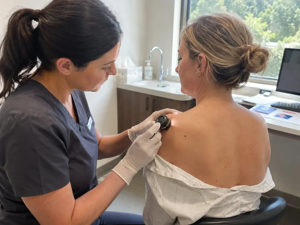
In the clinical dermatology and aesthetic medicine sectors, understanding the cellular changes of a skin lesion post-intervention is essential. When people ask how frequently a mole can grow back after being removed, the reply varies solely with the method of removal and the depth of the original lesion. A mole re-growing is actually quite a common and normal biological phenomenon, especially when a benign nevus (mole) is being treated by superficial methods rather than full-thickness excision.
In biometric terms, a mole consists of tightly grouped melanocytes (pigment-producing cells) that can infiltrate the deeper skin layers. If even a very small cluster of these cells is inadvertently left behind in the tissue during the procedure, those cells still have the full capability to proliferate. After several weeks or months, these cells move back up to the surface of the skin, leading to the pigmentation coming back in or near the area of the scar.
The Comparison Of Shave Excision Versus Full-Thickness Removal
The chances of a mole growing back are closely linked to the specific surgical approach that your surgeon decides to take. A shave excision is a highly favored method for raised, cosmetic moles because it uses a small blade to shave the lesion off level with the skin surface, resulting in a very low-profile scar. However, since this method intentionally does not disrupt the tissue very deeply, it often leaves the mole’s dermal root completely in the skin, which may lead to the mole returning at a rate of fifteen to twenty percent over time.
On the other hand, a full-thickness punch or surgical ellipse excision gives a certain amount of clearance at the microscopic level. The doctor removes the dermal tissue completely, including a small rim of unaltered tissue, while taking out the mole’s deep root. They then close the wound with high-tech, very fine stitches. This method does require the tissue to go through a longer and more involved healing period. However, it brings the melanocyte recurrence almost to zero, giving a very permanent solution to deep or persistent lesions.
Keeping The Skin a Perfect Canvas to Showcase Your Athletic Figure
One of the most important things about skin management after mole removal is to appreciate how a flawless skin canvas acts as a direct reflection of your overall physicality and long-term architectural goals. A person who is focused on body sculpting and especially those who are into gluteal and lower body hypertrophy for a striking hourglass figure, will find dermal appearance very influential. A large, dark, or recurrent mole situated along the most visible lines, like collarbones, shoulders, or midsection, can be so much of a visual distraction as to break the entire flow of beautiful muscle definition.
By applying the mathematically calibrated surgical removal methods to permanently clear these localized skin imperfections, the doctor dramatically purifies the skin architecture. Removal of a recurring lesion guarantees that your skin will be as smooth, uniform, and high definition as your physical frame, allowing natural bone structure and narrow waistline to feature prominently without any distracting visual interruptions. Paying such close attention to systemic harmony leads to the upper body aesthetics conveying the same uncompromised discipline and refined vitality as the physical proportions.
Differentiating Benign Recurrence From Changes
It is very important for patients to understand that a mole’s growth is not always a simple issue. In fact, it is a visual development that necessitates close examination by a doctor. A recurring nevus, commonly known as a pseudomelanoma, usually looks like a tiny dot of brown or black pigment within the boundaries of the healing surgical scar. Although this pigmentation sometimes looks uneven or even alarming, it is almost always simply a benign biological response of the remaining healthy cells to the environment of the new scar tissue.
Nevertheless, since the appearance of a benign recurrence can sometimes resemble the irregular borders and asymmetrical pigmentation seen in atypical lesions or early melanomas, it would be dangerous to rely on guessing. For safety reasons, any mole that shows growth after a previous removal should be subjected to high-definition dermoscopy for a detailed examination. If the pigment pattern shows any irregularity, the expert surgical team will perform an immediate and thorough secondary biopsy in order to tease out the exact nature of the lesion and to put your mind at the highest level of ease.
Mole Removal in Turkey
At LIN Europe Clinic, you will find a premier global medical paradise where your personal structural and dermal transformation will be handled with great clinical skill and deep empathetic care. We understand that even simple dermatological surgeries deserve a sophisticated, transparent, and very personalized setting that puts evidence-based medicine above everything else. LIN Europe Clinic in Turkey is known worldwide for the expertise of advanced facial and body contouring and is a calm environment where your treatment plan will be determined by the highest safety standards for patients.
When you trust our Istanbul team of specialists, you will be part of the elite medical framework that not only analyzes your exact skin chemistry but also lesion depth very carefully before deciding on a surgical plan. To guarantee that cosmetic results will be beautified and the risk of recurrence minimized, our diagnostic and closure techniques are world-class and tension-free. Come to LIN Europe Clinic for highly professional and comprehensive care where your beautiful and smooth balance is our mission, safely and skillfully, in the heart of Turkey.
FAQ:
Sometimes, a mole can grow again if a few pigment cells that cannot be seen with the naked eye remain in the skin after the operation. This is the most probable result after a superficial shave excision, which removes only the top part of the spotted lesion but leaves the deep dermal root intact.
One of the best methods to prevent the return of the mole is the full-thickness surgical excision. This is a sophisticated technique that completely removes a lesion and its roots deeply embedded in the skin, which will reduce the chances of the mole coming back to almost zero.
Reappearance of dark or brown spots in your healing wound is quite a common thing to happen. However, you should not hesitate to consult a specialist immediately. He/she is going to perform a high-definition dermoscopy evaluation of the scar in order to confirm that it is merely a benign re-occurrence of the mole rather than an atypical transformation.
In most cases, a mole that grows back is what is known as a pseudomelanoma. This is simply a healthy group of pigment cells that are multiplying in a new scar and is not cancerous. On the other hand, one has to see a professional for a clinical examination so that any abnormalities can be ruled out.
When you want a mole removed, you would want to combine your procedure with a visit to a dermatology team that is second to none internationally, as well as keen proficiency in skin evaluation by surgical mapping. LIN Europe Clinic in Turkey is staffed by a team of very knowledgeable medical physicians or a highly trained medical team which is able to design each treatment in such a way that the patient will get a totally smooth skin and will be free of blemishes, yet with the least possible scars.
Cockereell, C. J. (2002). Diagnostic Histopathology of Benign and Malignant Melanocytic Lesions. McGraw-Hill.
Goldberg, L. H., & Kim, J. Y. (2005). The shave excision technique for benign dermatological lesions: outcomes and recurrence rates. Journal of the American Academy of Dermatology, 53(4), 644-649.
Requena, L., & Kiryu, H. (1995). Pseudomelanoma: a review of recurring melanocytic nevi following incomplete surgical removal. American Journal of Dermatopathology, 17(2), 181-189.