Reshapes and repositions prominent ears.
The Anatomical Foundations and Pre-Surgical Cartilage Mapping
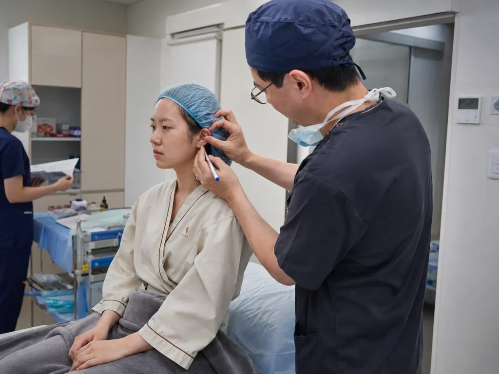
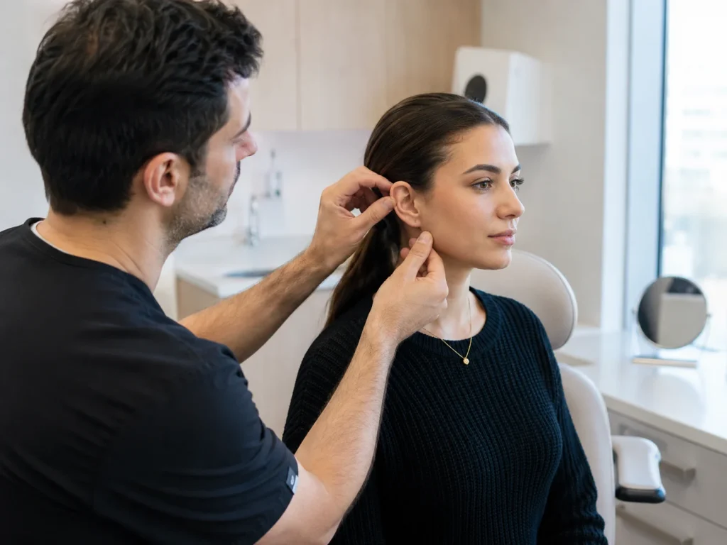
In the very specialized field of facial plastic surgery and aesthetic reconstruction, where one aims at natural cranial proportions very strictly, compliance with absolute structural frameworks is non-negotiable. Otoplasty, or more colloquially, ear reshaping or ear pinning, is a correction method for prominent, uneven ears or ears with structural problems. Preparation of the operation starts with a detailed anatomical determination of the auricle, mainly the antihelix fold, conchal bowl depth, and lobule angle.
The main concern at the time of creation of the plan is to spot exactly the biomechanical reasons for protrusion or deformation. If the antihelix fold is not properly formed, then the upper part of the ear will stick out. On the other hand, a large conchal bowl will push the whole ear away from the head. Surgeons go beyond just pictures; they also use vector analysis to figure out exactly how many degrees and which way the ear needs to be rotated and folded. Such a preparation based on mathematical formulas guarantees that final changes will be the ones that fit the patient’s natural facial profiles and that a right balance between cosmetic correction and structural integrity will be maintained.
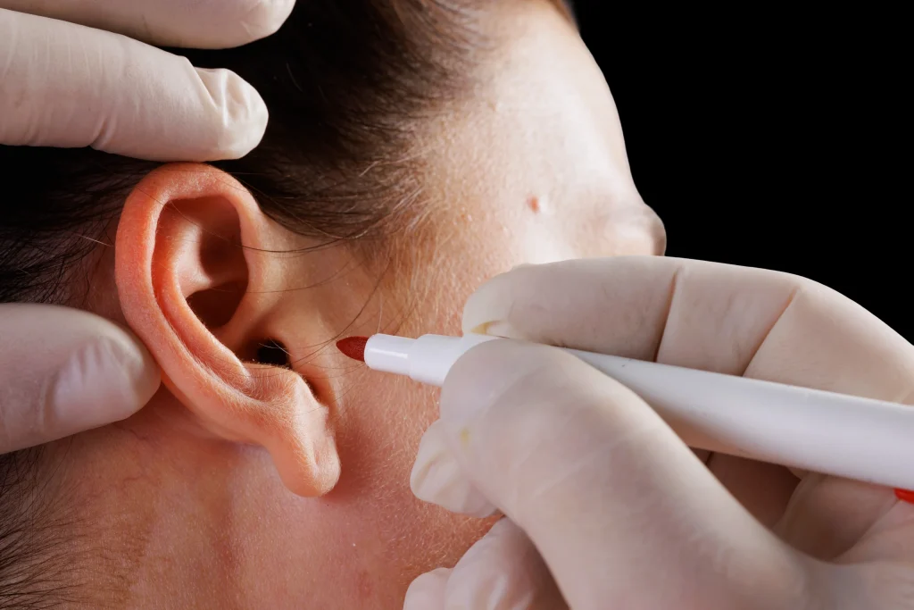
Intraoperative Mechanics: Anesthesia, Incisions, and Cartilage Sculpting
Otoplasty can be defined as a delicate tissue handling, and nowadays it is commonly done as a one-day procedure. For adults, doctors usually use local anesthesia combined with intravenous sedation, which renders the patient painless, while for children, general anesthesia is given, which makes the patient completely immobile. In the case of optimal anesthesia, the physical change will start with the very careful and well-thought-out incisional placement.
It is just a thin vertical cut the surgeon makes, but it goes only through the back side of the ear and is totally unnoticeable because it is located within the natural skin fold where the ear meets the skull. Through this micro-exposure, the underlying auricular cartilage is carefully access-mapped. Depending on the patient’s unique anatomical blueprint, the surgeon employs a combination of cartilage scoring—which alters the inherent spring and memory of the tissue—and permanent internal sutures (such as Mustarde or Furnas stitches). These specialized matrix sutures act as an internal scaffolding, folding the cartilage backward to create a sharp antihelical fold or anchoring the conchal cartilage directly to the mastoid fascia to reduce projection without sacrificing natural ear flexibility.
Preserving Post-Surgical Microcirculation
Strictly following recovery boundaries is a must for those who lead a disciplined lifestyle and want perfection in their appearance. People for whom physical exercise, a balanced body, and a neat face are very important will need to be very careful with their ears, especially during the immediate post-operative period. Ears are likely to remain irritated internally when there is trauma, friction, or sudden pressure. Besides being a strict biological penalty, it is also likely to cause disruption of internal sutures, formation of hematomas as well as alteration of a delicate blood supply that is a must for tissue survival.
Structured recovery plans will not only help you to save your body’s cellular environment but also improve your structural investment. It’s a fact that after the surgery, the microcirculation of the skin of the ear area changes drastically, and so avoidance of having to put clothes over the head, as well as resting in a supine position, will be the measures that help in preventing fluid from getting badly pooled. Such diligence in care ensures that the freshly folded cartilage gets a steady flow of oxygenated blood without any disruption, which results in your fresh facial contours, symmetric placement of the ears, and balanced silhouette healing with complete structural efficiency.
Post-Operative Behavioral Modifications
Your post-operative behavior will be your strongest tool not only to maintain technically successful surgery but also to help your new cartilage shape be preserved in the long run. The battle for retention of that new positional form happens not only within the tissues, but also as the patient interacts with his/her surroundings. If these new shapes are bent or pulled when a patient is sleeping, the internal retention sutures can tear through the soft cartilage matrix, leading to a recurrence of the protrusion.
Some of the very efficient behavioral methods that help post-surgical recovery also include the use of compression dressings. Patients are advised to wear a thick bandage that is able to provide support to their ears, always and uninterruptedly, for the first week. This is done to reduce swelling and keep the ears immobilized. Removal of this wrap should be followed by wearing a soft, stretchy headband 24/7 for another two weeks and then using it only at night for an additional month. This kind of non-invasive mechanical barrier free of all gravitational and frictional influences during sleep guarantees that ears stay perfectly flat against the cranial framework while the internal scar’s primary maturation is taking place.
Otoplasty in Turkey
LIN Europe Clinic is a great choice if you want to get treated in a world-class medical center where your facial aesthetics, body transformations, and surgical pathways are handled with the highest expertise and full empathy. We understand that healing from a powerful surgical procedure on the face is difficult, and therefore, you will need a highly sophisticated, transparent, and very supportive environment that is totally based on evidence-based medicine. At LIN Europe Clinic in Turkey, you will find international-standard advanced facial remodeling and cosmetic aftercare, accompanied by patient safety as a major concern at all times.
By choosing us at the LIN Europe Clinic in Istanbul, you are in very good hands. Specialists will monitor recovery parameters with decent diagnostic tools all the time for your peace of mind. Step-by-step through every phase of tissue maturation and lifestyle integration guidance coming postoperatively to each individual personally, and healing along the way is what we provide. Your cosmetic investment and systemic health will be safeguarded perfectly by our excellent medical personnel, while you will be free to enjoy your dream silhouette in a state of complete peace of mind. LIN Europe Clinic is your ticket to the refined, comprehensive care performed professionally and safely in the heart of Turkey.
FAQ:
Absolutely not. All otoplasty procedure’s incisions are made within the deepest natural fold behind the ears only. This well-planned placement ensures that after healing is complete, the scar will not be visible at all, neither from the front nor from the side.
Generally, after removing the surgical dressing, most patients might be willing and able to return to their work or school that does not require physical exertion within 5 to 7 days of the procedure. Nonetheless, to safeguard the healing ears, one should avoid engaging in exercise, heavy work, and contact sports for at least 4 to 6 weeks.
Indeed, otoplasty results are meant to last for a lifetime. At first, the cartilage is kept in its new position by internal permanent sutures, but over the first few months, the body generates strong internal scar tissue, which permanently secures the altered cartilage structure.
Certainly, it is one of the most frequently done surgical operation for children. A child needs to have ear cartilage size of about 85-90% of an adult before undergoing otoplasty, which is usually 5 or 6 years of age. Having surgery at this age would most likely be able to help a child prevent getting upset over the way other children treat him/her around that time.
After bending or pulling your ear by inadvertence during the sleep, you ought to watch for any bleeding, sudden very much swelling, or change in the ear’s position. Do not hesitate to get in touch with your surgical team so that together you can verify the condition of the internal retention sutures.
Mustardé, J. C. (1963). The correction of prominent ears using simple inverted mattress sutures. British Journal of Plastic Surgery, 16, 170-178.
Furnas, D. W. (1968). Correction of prominent ears by concha-mastoid sutures. Plastic and Reconstructive Surgery, 42(3), 189-193.
Elliott, R. A. (1990). Otoplasty: A review of surgical techniques and long-term architectural stability. Aesthetic Plastic Surgery, 14(1), 25-31.