Yes, recurrent moles can be removed again.
The Cellular Structure Of Recurrent Melanocytic Nevi
In clinical dermatology and aesthetic medicine, secondary interventions on initially treated skin lesions are a common clinical practice. The answer to whether a mole removal procedure can be safely repeated, from a biological standpoint, is an unequivocal yes. A benign nevus can sometimes regenerate inside the margins of a healing surgical scar, and this is exactly why a secondary operation is needed. Such a situation is a physiological phenomenon that occurs when small groups of pigment-producing melanocytes that have been left behind after the initial operation multiply and grow in the surgical scar area.
In terms of biometric analysis, a mole normally has very deep intradermal roots going down to layers that are well below the epidermis. If the first treatment was done in a superficial manner—say, with a cosmetic shave excision or electrosurgical ablation—the upper part of the mole will be successfully removed, but its deep cellular base will remain intact in the tissue bed. During several months, these leftover cell nests will proliferate and move themselves to the surface through the developing scar tissue, leading to a localized pigment recurrence that has to be removed again.
Guidelines For Surgical Clearance of Second-time Excision
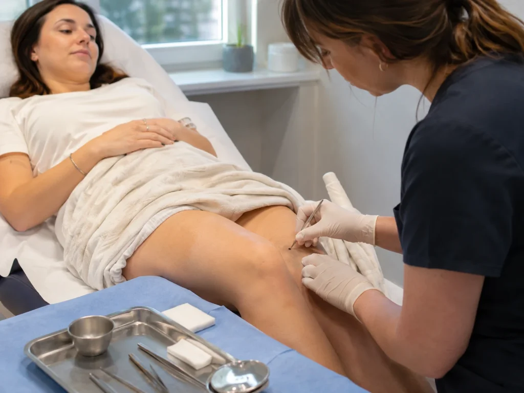
It can be challenging to perform a second removal on the very same anatomical location due to the presence of a pre-existing scar and changed dermal planes. Furthermore, it is usually recommended not to use the same superficial shave technique when dealing with a recurrent mole for a second time because it significantly increases the chances of a third recurrence. What a reconstructive specialist will do is resort to a definite full-thickness surgical ellipse or punch excision to completely get rid of the lesion.
This extensive secondary procedure involves the surgeon making a full-thickness cut through the dermal framework and a slight extension of the incision margins beyond the previous scar tissue. As a result, it is guaranteed that the deepest root of the mole, along with any active melanocyte nests, will be completely removed from the tissue bed. The surgeon will then close the wound using skillfully done, multi-layered micro-sutures, which minimizes the possibility of a second recurrence practically to zero while at the same time promoting an excellent final cosmetic outcome of the skin canvas.
Keeping Your Dermal Canvas Clean To Support An Athletic Frame
A major factor in the correct handling of recurring skin blemishes is an understanding of how an immaculate, thoroughly even skin texture goes hand in hand with your overall body proportions and your fitness goals over the long term. Those who apply tremendous self-discipline and determination to building an athletic body—especially those who seriously target their lower body and glutes for development to achieve a perfect hourglass shape—will find that the quality of the skin visually is hugely influential. A dark blemish that keeps appearing will be highly noticeable, for example, on the neck, shoulders, or midsection, and it will not just be a visual disturbance but one that breaks the harmonious flow of your sculpted muscles.
By resorting to a thorough, full-thickness secondary excision for the resolute removal of this spot, the expert effectively restores the tissue framework of your skin. Complete restoration through removal of the pigment that has been continually recurring means that your skin canvas will be just as clean, high-definition, and evenly toned as your body, thus making your natural bone and narrow waistline visible in a most striking manner. Such serious system-wide consideration guarantees that your upper body can aesthetically communicate the same unyielding call for discipline and authentic health as is shown by your physical proportions.
Meticulous Diagnostics And Histopathological Confirmation
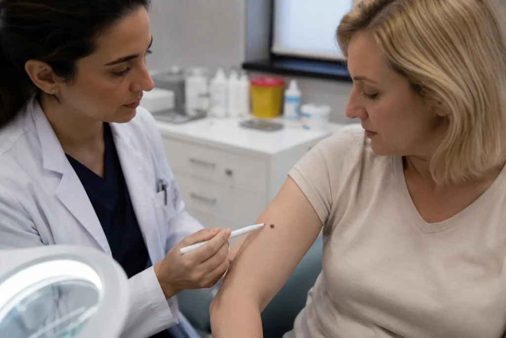
Absolute professional safety in the first place requires that patients be made aware that any mole showing signs of recurrence after an initial removal operation must be strictly diagnostically confirmed. Although most recurrence within a scar is caused by benign lesions—a condition called pseudomelanoma—certain visual clues, such as irregular borders and multiple colors in which the pigment presents upon recurrence, can cause concern as they are very similar to the signs of atypical or early malignant changes.
In the first place, ensuring 100% clinical safety means that there can be no room for guessing. No leading medical group would consider simply destroying or discarding a recurring lesion. Once the secondary full-thickness removal has been done, the excised tissue is sent without delay to a specialist dermatopathology laboratory for the formal histopathological study. The microscopic evaluation reveals the cellular makeup of the tissue and confirms that there is no lesion left at the margins, thereby giving you long-lasting peace of mind with respect to your skin health.
Treatment of Moles in Turkey
Opting for LIN Europe Clinic means stepping into a world-class medical oasis where you will be uniquely transformed structurally and dermally with clinical excellence and caring empathy at the highest level. We understand very well that re-treating a recurring skin lesion for the second time requires an extremely well-educated, transparent, and highly individualistic setting that makes evidence-based medicine the first priority, above all else. Located in a beautiful part of Turkey, LIN Europe Clinic is recognized worldwide for excellence in advanced facial and body contouring and, at the same time, offers a peaceful environment where you can be guided on your treatment path strictly by the highest patient safety standards.
Secondly, by putting your profound trust in our team of experts at LIN Europe Clinic in Istanbul, your second skin procedure will be orchestrated with the highest degree of accuracy. We combine world-class diagnostics, the use of high-resolution dermoscopy, and state-of-the-art tension-free internal suturing techniques to ensure that your secondary mole removal not only enhances your aesthetic appearance but also completely removes the underlying cellular roots. Discover the polished and all-around cared-for experience at LIN Europe Clinic and get your smoothly beautiful and flawlessly proportioned canvas, safely and expertly created in Turkey.
FAQ:
Yes, that is absolutely safe and quite normal, in fact, to have a mole taken off a second time in the event it starts growing back. Usually, a follow-up removal is needed only when pigment cells that were left behind after the first operation multiply inside the newly formed scar tissue.
Moles get back because the first treatment left very small groups of alive pigment cells deep within the skin. This happens very often after surface shave removals that cut the top part of the spot but leave the deeper portions untouched.
Dermatologists, to make sure the spot is permanently removed, will do a complete thick surgical removal during the 2nd visit. The process is to surgically remove the entire scar pocket and deep roots before the skin is closed with fine micro sutures.
Most moles that come back are just harmless growths called pseudomeleanomas. The only problem is that the pigment in a returning mole can look quite irregular, so a doctor has to examine the spot under a dermoscope and the tissue is then sent to a laboratory for formal testing.
A second complete excision will be a little longer than the first superficial one in order to fully clear the scar’s pocket, but precise, layered suturing techniques used will help the remaining line to heal as a flat, barely visible mark.
Cockereell, C. J. (2002). Diagnostic Histopathology of Benign and Malignant Melanocytic Lesions. McGraw-Hill.
Goldberg, L. H., & Kim, J. Y. (2005). The shave excision technique for benign dermatological lesions: outcomes and recurrence rates. Journal of the American Academy of Dermatology, 53(4), 644-649.
Requena, L., & Kiryu, H. (1995). Pseudomelanoma: a review of recurring melanocytic nevi following incomplete surgical removal. American Journal of Dermatopathology, 17(2), 181-189.